This package has moved
cellfinder-napari has merged with it's backend code and is now available as a single package called cellfinder.
We recommend you uninstall cellfinder-napari and instead use the functionality provided in the cellfinder package.
These changes are part of our wider restructuring of the BrainGlobe suite of tools and analysis pipelines, which you can keep up to date with on our blog.
cellfinder-napari


![]()


Efficient cell detection in large images (e.g. whole mouse brain images)
cellfinder-napari is a front-end to cellfinder-core to allow ease of use within the napari multidimensional image viewer. For more details on this approach, please see Tyson, Rousseau & Niedworok et al. (2021). This algorithm can also be used within the original
cellfinder software for
whole-brain microscopy analysis.
cellfinder-napari, cellfinder and cellfinder-core were developed by Charly Rousseau and Adam Tyson in the Margrie Lab, based on previous work by Christian Niedworok, generously supported by the Sainsbury Wellcome Centre.
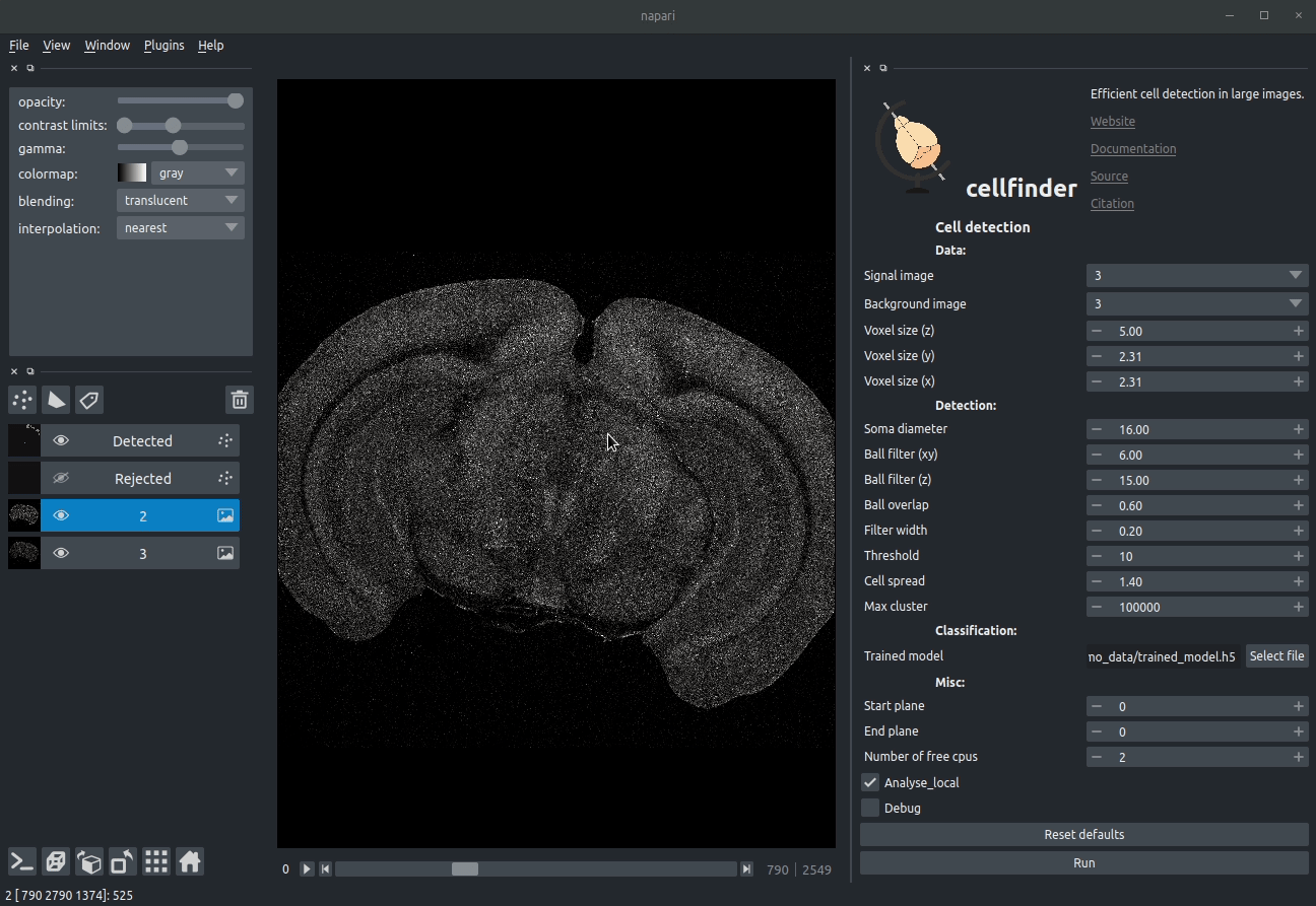
Visualising detected cells in the cellfinder napari plugin
Instructions
Installation
Once you have installed napari. You can install napari either through the napari plugin installation tool, or directly from PyPI with:
pip install cellfinder-napariUsage
Full documentation can be found here.
This software is at a very early stage, and was written with our data in mind. Over time we hope to support other data types/formats. If you have any questions or issues, please get in touch on the forum or by raising an issue.
Illustration
Introduction
cellfinder takes a stitched, but otherwise raw dataset with at least two channels:
- Background channel (i.e. autofluorescence)
- Signal channel, the one with the cells to be detected:
 Raw coronal serial two-photon mouse brain image showing labelled cells
Cell candidate detection
Classical image analysis (e.g. filters, thresholding) is used to find cell-like objects (with false positives):
 Candidate cells (including many artefacts)
Cell candidate classification
A deep-learning network (ResNet) is used to classify cell candidates as true cells or artefacts:
 Cassified cell candidates. Yellow - cells, Blue - artefacts
Contributing
Contributions to cellfinder-napari are more than welcome. Please see the developers guide.
Citing cellfinder
If you find this plugin useful, and use it in your research, please cite the paper outlining the cell detection algorithm:
Tyson, A. L., Rousseau, C. V., Niedworok, C. J., Keshavarzi, S., Tsitoura, C., Cossell, L., Strom, M. and Margrie, T. W. (2021) “A deep learning algorithm for 3D cell detection in whole mouse brain image datasets’ PLOS Computational Biology, 17(5), e1009074 https://doi.org/10.1371/journal.pcbi.1009074
If you use this, or any other tools in the brainglobe suite, please let us know, and we'd be happy to promote your paper/talk etc.


![pre-commit-ci[bot]](https://avatars.githubusercontent.com/u/66853113?size=120)


